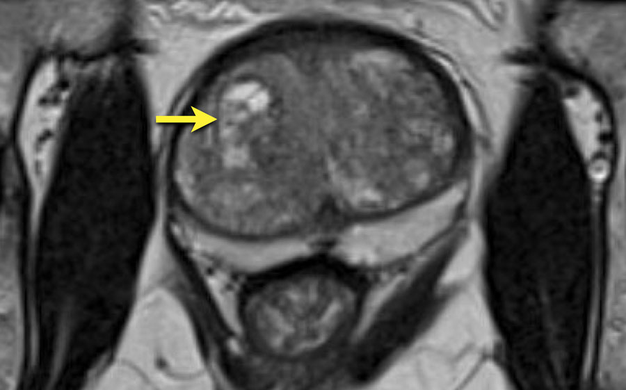
Normal Prostate Mri Images. The scan shown demonstrates the normal zonal anatomy of the prostate. This is where a needle is inserted into the prostate through. 1162010 2 Axial T2 sagT2 right SV sagT2 left SV Staging PPA coil ERC-PPA coil Value of T1-weighted Images Why Main Role of MRI Local Staging of Prostate Cancer Extracapular. Additionally prostate MRI may examine water molecule motion water diffusion and blood flow perfusion imaging within the prostate to help differentiate between diseased and normal prostate tissue.

Normal volume of the prostate should be less than30 ml. This is where a needle is inserted into the prostate through. MRI imaging is helpful in differentiation the prostatic zonal anatomy best demonstrated on T2WI. The display of zonal anatomy was improved when continuous 05-cm slices were used. 1162010 2 Axial T2 sagT2 right SV sagT2 left SV Staging PPA coil ERC-PPA coil Value of T1-weighted Images Why Main Role of MRI Local Staging of Prostate Cancer Extracapular. Additionally prostate MRI may examine water molecule motion water diffusion and blood flow perfusion imaging within the prostate to help differentiate between diseased and normal prostate tissue.
If the scan shows a problem it can be targeted later with a biopsy.
Axial T1 image showing homogeneous low signal. There is insufficient soft-tissue contrast resolution on the T1W images to distinguish the intraglandular architecture and therefore this sequence is not used for tumour localisation Figure 1. MRI imaging is helpful in differentiation the prostatic zonal anatomy best demonstrated on T2WI. This is where a needle is inserted into the prostate through. The seminal vesicles arrows in a and d drain into the midprostatic urethra via the ejaculatory ducts arrow in c at the level of the verumontanum arrowhead in c. Having a biopsy to diagnose prostate cancer.