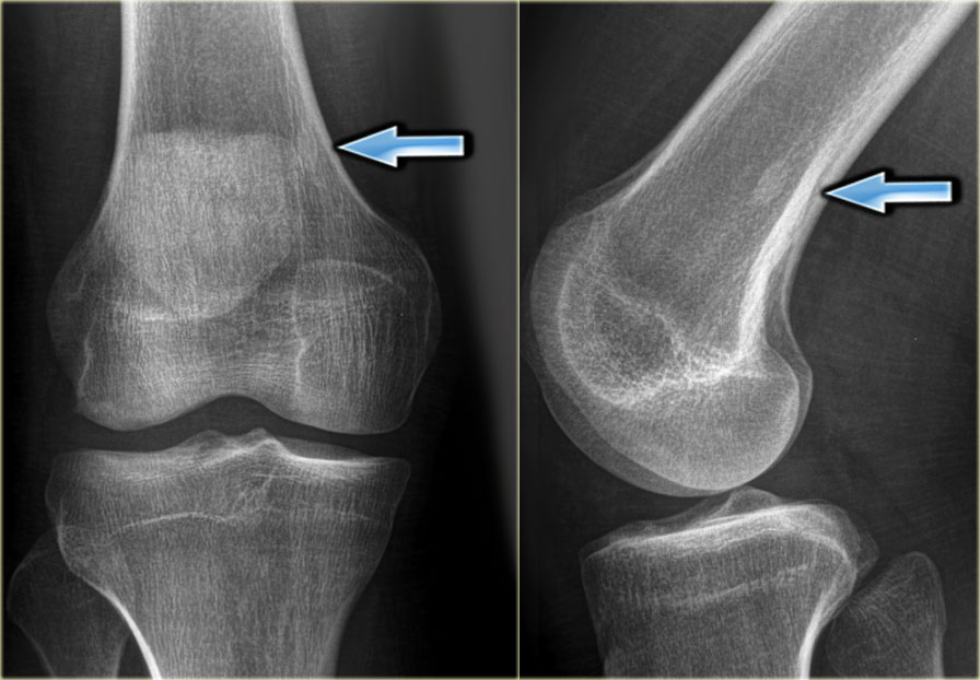
Lytic Or Sclerotic Lesion. In pediatric patients 3. Typically presents as a lytic lesion in a flat bone vertebra or diaphysis of long bone. May show extinction and become sclerotic and indolent. Ask the patient or the clinician about this.

The sclerosis is caused by increased calcium deposits forming in a small area of he bone. Mucinous adenocarcinoma of the gastrointestinal tract eg. Appear lytic on plain films on T2W images heteroge-. Patients with sclerotic lesions due to metastasis often have a history of prior malignant disease. May present as well-defined osteolytic ill-defined osteolytic and also as sclerotic bone lesion. Must be considered in the differential diagnosis of any bone lesion in a patient 40 years.
MR usually shows a large amount of reactive changes in bone and soft tissue.
Sclerotic lesions presenting as focalmultifocal lesions on MRI include bone infarct chronic granulomatous or fungal infections bone island and various primary as well as secondary neoplasms. By knowing the typical behavior of the metastatic lesion - lytic or blastic - you can help sort between the types to make the mnemonic even more useful. Kidney thyroid lung lytic induces bone destruction. In the majority of skeletal metastases new bone develops simultaneously with bone destruction. The roentgenogram indicates the net effect of these two processes. May show extinction and become sclerotic and indolent.