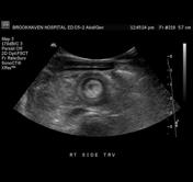
Bulls Eye Sign Ultrasound. A tri-laminar appearance is also seen in larger cysts. The pathology reports of those lesions biopsied were reviewed. A typical ultrasound phenotype of hepatic candidiasis is the bulls eye or target pattern with a peripheral hypoechoic halo encircling a central hyperechoic core Fig. The bulls-eye sign was found to be a specific indicator of normal hematopoietic marrow sensitivity 95.

Bulls eye appearance is a common presentation of metastases from gastrointestinal GI adenocarcinomas and represents histological findings that show an area of. There are many bulls eye signs also referred to as target signs. 89 Occasionally hepatosplenic candidiasis may not be detectable with B-mode ultrasound. After IV contrast enhancement the veins and fibrous tissue can accumulate contrast agent more intensively than the tissue of regenerative nodes thus forming the bulls eye sign. When demonstrated by ultrasound this finding should suggest portal venous hypertension. The bulls eye sign is seen in a spectrum of benign cystic lesions that include cysts and fibrocystic change.
The pathology reports of those lesions biopsied were reviewed.
A retrospective review of 383 patients having an EI for diagnostic breast ultrasound evaluation was reviewed. A new toothpaste sign is proposed to suggest low grade DCIS in a lesion with elastographic ratios of more than 1. Doctors in 147 specialties are here to answer your questions or. It was not visualized in any patients with cholelithiasis who had no evidence of cirrhosis. A typical ultrasound phenotype of hepatic candidiasis is the bulls eye or target pattern with a peripheral hypoechoic halo encircling a central hyperechoic core Fig. Earliest lesion seen in Crohn disease gastric lymphoma with central ulceration 4.